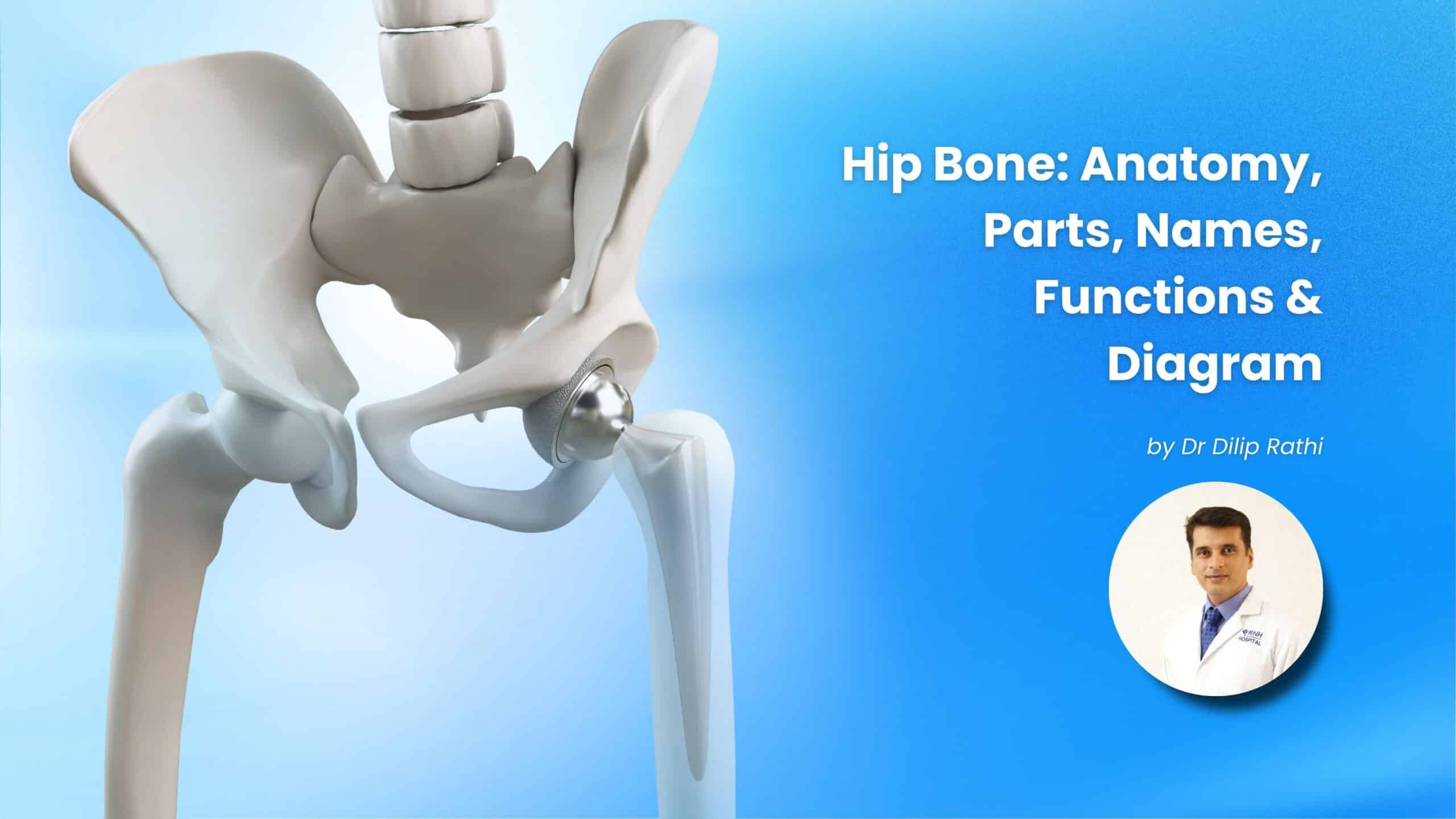
Hip Bone: Anatomy, Parts, Names, Functions & Diagram
By Dr Dilip Rathi, M.B.B.S, MS. (Ortho), Joint Replacement & Trauma Specialist (30+ years of experience)
The hip bone plays an essential role in supporting the body, maintaining balance, and enabling movements such as walking, sitting, standing, and running. Understanding hip bone anatomy can help you recognize the importance of hip health and identify symptoms that may require medical attention.
The hip bone is part of the pelvis and connects with the thigh bone to form the hip joint. Problems such as arthritis, fractures, hip dislocation, and muscle or tendon injuries can cause pain around this region.
At RNH Hospital, Nagpur, understanding the causes of hip pain is an important step toward appropriate orthopedic evaluation and treatment. In this article, we explain what the hip bone is, where it is located, its parts and functions, and the common reasons for hip bone pain.
What Is Hip Bone?
The hip bone is a large, irregularly shaped bone that forms part of the pelvic skeleton. In adults, each hip bone develops from three bones that fuse together: the ilium, ischium, and pubis.
There are two hip bones in the human body, one on the left side and one on the right side. Together with the sacrum and coccyx, they form the bony pelvis, which supports the upper body and protects organs in the pelvic region.
Each hip bone contains a cup-shaped structure called the acetabulum. This socket holds the rounded head of the femur, or thigh bone, forming the hip joint.
The hip joint is a ball-and-socket joint that allows a wide range of movements while providing stability and supporting body weight.
Where Is the Hip Bone Located?
The hip bones are located on both sides of the lower torso, between the abdomen and the upper thighs. They form the left and right sides of the pelvis.
You can generally feel parts of the hip bone around the sides of your waist, near the bony prominences commonly called the hip points. The exact location of discomfort may help a healthcare professional identify whether the problem involves the bone, hip joint, muscles, tendons, or surrounding structures.
The hip region includes several important structures:
- Pelvis: The bony framework that supports the body and protects pelvic organs.
- Hip joint: The joint connecting the pelvis to the femur.
- Femur: The thigh bone, whose upper end fits into the hip socket.
- Muscles and tendons: Structures that support the hip and help control movement.
- Ligaments: Strong connective tissues that help stabilize the joint.
If you experience persistent pain around the hip, an orthopedic specialist can assess the location, severity, and possible cause.
Hip Bone Anatomy: Understanding Its Structure
Hip bone anatomy consists of three main parts: the ilium, ischium, and pubis. These parts fuse during development to form a single hip bone on each side of the pelvis.
1. Ilium
The ilium is the largest and uppermost part of the hip bone. It forms the broad, wing-shaped region of the pelvis that can be felt around the waist.
Functions of the ilium:
- Supports the upper part of the pelvis.
- Provides attachment points for abdominal, back, and hip muscles.
- Helps distribute body weight through the pelvic skeleton.
2. Ischium
The ischium forms the lower and back portion of the hip bone. Its prominent lower region, called the ischial tuberosity, supports much of the body’s weight when sitting.
Functions of the ischium:
- Provides support while sitting.
- Serves as an attachment point for muscles and ligaments.
- Contributes to the structure and stability of the pelvis.
3. Pubis
The pubis is the front portion of the hip bone. The left and right pubic bones meet at a joint called the pubic symphysis.
Functions of the pubis:
- Forms the front portion of the pelvic ring.
- Provides attachment points for muscles of the abdomen and inner thigh.
- Helps maintain pelvic stability.
These three parts meet around the acetabulum, the socket that forms part of the hip joint.
Hip Bone Parts and Their Names
The hip bone contains several important anatomical landmarks in addition to the three main parts.
| Hip bone part | Location and function |
| Ilium | Upper, broad part of the hip bone |
| Ischium | Lower and back portion; supports the body while sitting |
| Pubis | Front portion of the hip bone |
| Acetabulum | Socket that receives the head of the femur |
| Iliac crest | Upper border of the ilium |
| Ischial tuberosity | Bony prominence that supports sitting |
| Pubic symphysis | Joint connecting the left and right pubic bones at the front |
Knowing these hip bone parts helps explain how the pelvis provides stability while allowing movement at the hip joint.
Hip Bone Diagram: How to Understand the Structure
A labelled hip bone diagram typically shows the ilium, ischium, pubis, acetabulum, and important bony landmarks.
When viewing a hip bone diagram, look for the following structures:
- Ilium: The large, fan-shaped upper section.
- Ischium: The lower and posterior section.
- Pubis: The anterior section.
- Acetabulum: The socket on the outer side where the femoral head fits.
- Femoral head: The rounded upper end of the thigh bone that sits inside the socket.
The acetabulum and femoral head work together to form the hip joint. Cartilage covering the joint surfaces allows smooth movement, while ligaments, muscles, and tendons help stabilize the joint.
A labelled diagram is useful for understanding the relationship between the pelvic bones and the thigh bone, especially when learning about hip injuries or joint conditions.
What Is the Name of the Hip Bone?
The anatomical name for each hip bone is the coxal bone, also known as the os coxae or innominate bone. It is formed by the fusion of the ilium, ischium, and pubis.
The hip bone should not be confused with the femur. The femur is the thigh bone, and its rounded upper end fits into the acetabulum of the hip bone to form the hip joint.
Both bones are important for walking, standing, and supporting body weight.
What Are the Functions of the Hip Bone?
The hip bone performs several important functions that help maintain mobility and structural stability.
1. Supports Body Weight
The pelvic skeleton helps transfer body weight from the spine to the lower limbs during standing and walking.
2. Enables Movement
The hip bone forms the socket of the hip joint, allowing the thigh to move forward, backward, sideways, and rotationally.
3. Protects Internal Organs
The bony pelvis provides protection for organs within the pelvic cavity, including the urinary bladder and parts of the reproductive system.
4. Provides Muscle Attachment
Several muscles involved in walking, running, sitting, and maintaining posture attach to the hip bones.
5. Maintains Balance and Stability
The pelvis provides a stable foundation for the trunk and helps coordinate movements between the upper body and lower limbs.
Which Bone Comes Out of the Socket in Hip Dislocation?
In a hip dislocation, the head of the femur, or thigh bone, moves out of the acetabulum, the socket of the hip bone.
The hip joint is normally held together by strong ligaments, muscles, and its surrounding joint capsule. A significant force, such as a road traffic accident or a serious fall, can cause the femoral head to move out of its normal position.
Hip dislocation may cause:
- Sudden, severe hip or groin pain.
- Difficulty or inability to move the affected leg.
- An unusual position or appearance of the leg.
- Swelling and tenderness around the hip.
Hip dislocation is a medical emergency. If you suspect a dislocation, seek emergency medical care immediately. Do not try to push the joint back into place or move the injured leg unnecessarily. Prompt treatment is important to reduce the risk of complications, including damage to the blood supply of the femoral head.
Why Do Hip Bones Pain?
Hip bone pain can occur for several reasons. The location of the pain, the activities that trigger it, your age, and any history of injury can help a healthcare professional determine the likely cause.
Common causes include:
1. Arthritis
Osteoarthritis can damage the cartilage in the hip joint, leading to pain, stiffness, and reduced movement. Symptoms may worsen during walking or after prolonged activity.
2. Muscle or Tendon Injuries
Strains, overuse, and inflammation involving the muscles or tendons around the hip can cause pain during movement or exercise.
3. Hip Fractures
A fracture may occur after a fall, an accident, or a relatively minor injury in someone with weakened bones. Severe pain and difficulty standing or bearing weight may occur.
4. Hip Bursitis
Inflammation of a fluid-filled sac near the outer hip can cause pain and tenderness, particularly when lying on the affected side or walking.
5. Hip Dislocation
When the femoral head moves out of the acetabulum, it can cause severe pain and an inability to move the joint normally.
6. Referred Pain
Pain felt around the hip may sometimes originate from the lower back, nerves, or other nearby structures rather than the hip bone itself.
Persistent, worsening, or unexplained hip pain should be assessed by a healthcare professional. Sudden severe pain following an injury, an inability to bear weight, or a visibly deformed hip requires urgent medical attention.
How Are Hip Bone Problems Diagnosed?
An orthopedic specialist may assess the hip through a physical examination, medical history, and appropriate investigations.
Depending on the symptoms, diagnostic tests may include:
- X-ray: Helps identify fractures, joint alignment problems, and certain changes associated with arthritis.
- MRI: Provides detailed images of soft tissues, bone marrow, cartilage, and some injuries that may not be visible on an X-ray.
- CT scan: May help evaluate complex fractures or detailed bone anatomy.
Not every patient needs every test. The appropriate investigation depends on the suspected condition and clinical findings.
Treatment Options for Hip Bone Pain
Treatment depends on the underlying cause, the severity of symptoms, and the patient’s overall health.
Possible approaches include:
- Activity modification: Temporarily reducing activities that worsen symptoms.
- Medication: Appropriate pain-relieving or anti-inflammatory medication when recommended by a healthcare professional.
- Physiotherapy: Guided exercises to improve strength, flexibility, balance, and joint function when suitable.
- Fracture treatment: May involve immobilization, restricted weight-bearing, or surgery, depending on the fracture.
- Hip replacement surgery: May be considered for certain advanced joint conditions when other appropriate treatments no longer provide sufficient relief.
Avoid self-diagnosing persistent hip pain or starting exercises that aggravate your symptoms. An individualized treatment plan should be based on an accurate diagnosis.
When Should You Consult an Orthopedic Specialist?
Consider an orthopedic evaluation if you experience persistent hip pain, stiffness, reduced range of motion, discomfort while walking, or difficulty with everyday activities.
Seek urgent medical attention for severe pain after a fall or accident, inability to bear weight, suspected hip dislocation, or a sudden change in the shape or position of the leg.
At RNH Hospital, Nagpur, patients can seek orthopedic evaluation for hip and joint concerns. The appropriate treatment depends on the clinical assessment and the nature of the condition.
Early evaluation can help identify the cause of symptoms and guide suitable treatment.
Frequently Asked Questions About the Hip Bone
1. What is a hip bone?
The hip bone is a large bone that forms the left or right side of the pelvis. Each hip bone consists of three fused parts: the ilium, ischium, and pubis. It supports body weight and forms the socket of the hip joint.
2. Where is the hip bone located?
The hip bones are located on both sides of the lower torso, between the abdomen and the upper thighs. They form the sides of the pelvis and connect with the femurs through the hip joints.
3. Where is the hip bone in the human body?
The hip bone is part of the pelvic skeleton. You can feel some of its outer bony prominences near the sides of your waist. The hip joint itself is located deeper, where the femoral head fits into the acetabulum.
4. What are the three parts of the hip bone?
The three main parts are the ilium, ischium, and pubis. They fuse during development to form each adult hip bone.
5. What is the scientific name of the hip bone?
The anatomical name is the coxal bone, also called the os coxae or innominate bone.
6. Which bone comes out of the socket in hip dislocation?
The head of the femur, or thigh bone, comes out of the acetabulum, which is the socket in the hip bone. A suspected hip dislocation requires immediate emergency medical care.
7. Why do hip bones pain?
Hip-region pain can result from arthritis, muscle or tendon injuries, bursitis, fractures, hip dislocation, or referred pain from the lower back. The cause can be determined through clinical evaluation and, when necessary, imaging tests.
8. Can hip bone pain be treated without surgery?
Some causes of hip pain can improve with activity modification, medication, and physiotherapy. However, fractures, severe joint damage, and certain other conditions may require surgical treatment. The appropriate approach depends on the diagnosis.
9. Is the hip bone the same as the thigh bone?
No. The hip bone forms part of the pelvis, while the femur is the thigh bone. The head of the femur fits into the acetabulum of the hip bone to form the hip joint.
Conclusion
The hip bone is essential for supporting body weight, maintaining balance, protecting pelvic organs, and enabling movement. Understanding hip bone anatomy, its three main parts, and the structure of the hip joint can help you better understand common hip conditions.
If you experience persistent hip pain, stiffness, or difficulty walking, consult an orthopedic specialist for an appropriate evaluation. For sudden severe pain or a suspected hip dislocation or fracture, seek emergency care.
RNH Hospital, Nagpur, provides an orthopedic care setting for evaluating hip and joint concerns and helping patients understand suitable treatment options based on their individual needs.